Сentaur U - Scanning AFM/Confocal/Raman/Fluorescence system for Raman/Fluorescence and AFM/Raman (TERS) imaging
Centaur U - Scanning AFM/Confocal/Raman/Fluorescence system for Raman/Fluorescence and AFM/Raman (TERS) imaging
Centaur combines:
- Scanning Probe Microscope;
- Inverted or Upright Optical Microscope;
- Laser Confocal Microscope;
- Raman Confocal Microscope;
- Fluorescence Confocal Microscope.

Applications:
- Scanning Probe Microscopy;
- Raman Confocal Microscopy;
- Fluorescence Confocal Microscopy;
- Near-Field Scanning Microscopy;
- Tip-Enhanced Raman Spectroscopy (TERS);
- Tip-Enhanced Fluorescent Spectroscopy (TEFS).
Where to use:
- Chemistry. Combination of methods of scanning probe microscopy and Raman spectroscopy allows the analysis of the composition and structure of organic and inorganic substances, traditional and composite materials;
- Physics. Investigation of physical characteristics of surface and subsurface layers of substances and materials;
- Biology. Study of tissues, cells and their structures, biological molecules and the interactions between them;
- Interdisciplinary research. Research in the field of nanotechnology, pharmaceuticals, materials science, mineralogy, geology, forensic, analysis of art and many others.
")
Advantages of Centaur:
- Dual independent scanners (in head and base);
- Multiple simultaneous signal recording (confocal, spectra, topography, phase etc.);
- Full spectra recording in each scan point (hyperspectral imaging);
- Integration with virtually unmodified upright or inverted optical microscopes to work with transparent and none transparent samples;
- Modern cross-platform software (for all the Centaur units).

Components:
- Scanning Probe Microscope Certus;
- XY - sample piezo scanning stage Ratis;
- Confocal unit;
- Monochromator;
- Optical microscope (upright or inverted);
- Digital controller EG-3000;
- Software NSpec.
Raman spectra of carbon nanotubes (CNTs).
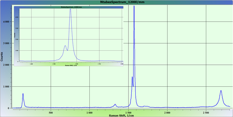
Raman spectra of carbon nanotubes (CNTs).
Grating 1200 lin/mm, exposition time 10 sec, monochromator pinhole 20 μm.
Demonstration of obtaining spectral information from surface with carbon nanotube.
Carbon nanotubes (CNTs) on glass surface. Confocal Raman image. One layer scanning.
Image size 25х25 μm. 100х100 points. Exposition time at point - 0,05 sec.
Images was obtained with AFM-Raman-Confocal system Centaur in confocal Raman microscope mode.
The sample was provided by Prof. V.K.Nevolin (www.nanotube.ru).